
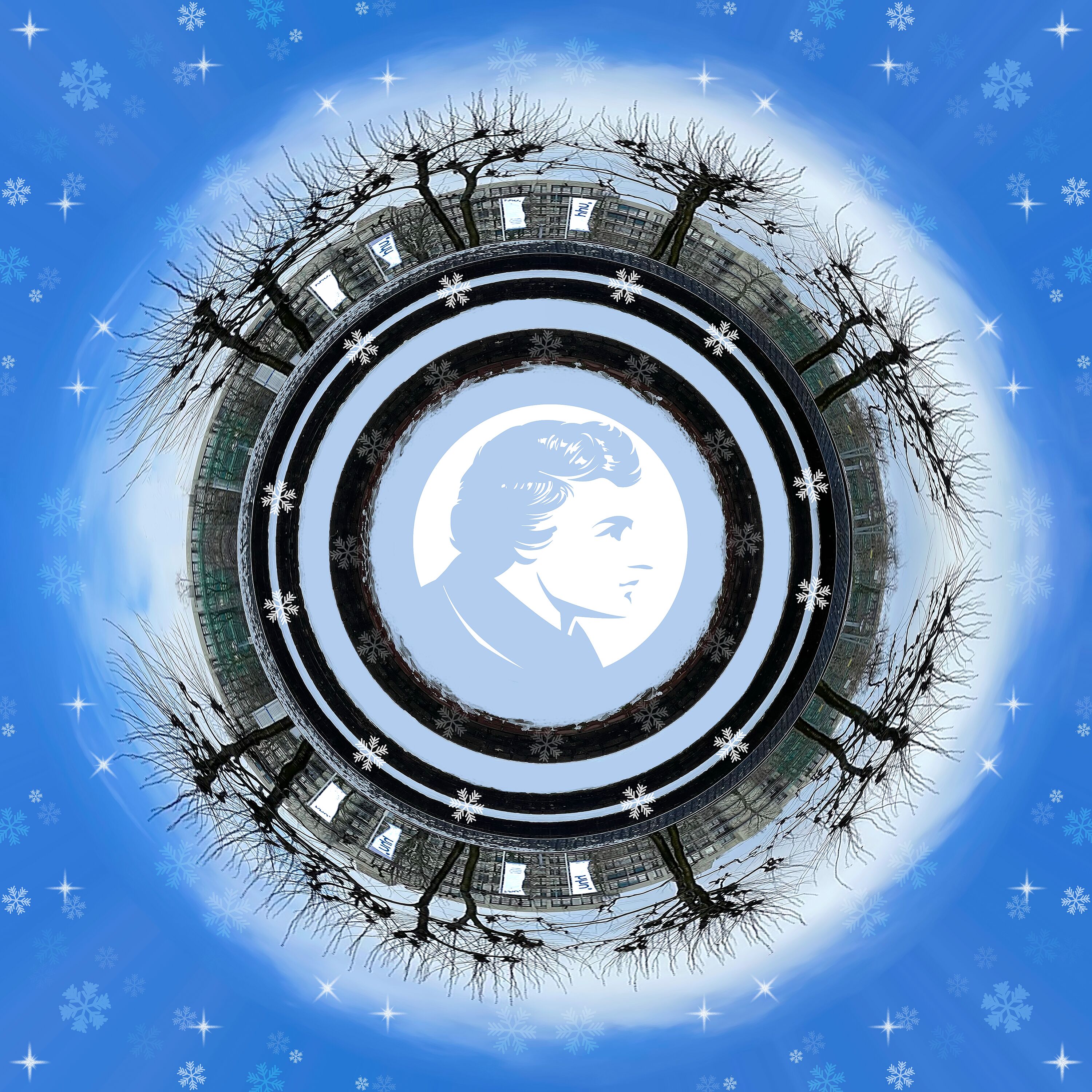
pART of Research - Calendar 2023
There is a lot of work in science - and also a lot of art. Whether it’s the coffee cup print on important notes, a picture from the last field research or impurities under the microscope - surprises, mishaps or the long-awaited result. And science is exchange.
This year 27 pictures from four of the five faculties were submitted to the competition under the motto: "Your Research - Your Art Work". We would like to thank all participants and all those who took the time to vote! The 12 most popular images (at least one from each of the participating faculties) are compiled in the "pART of Research Calendar 2023".
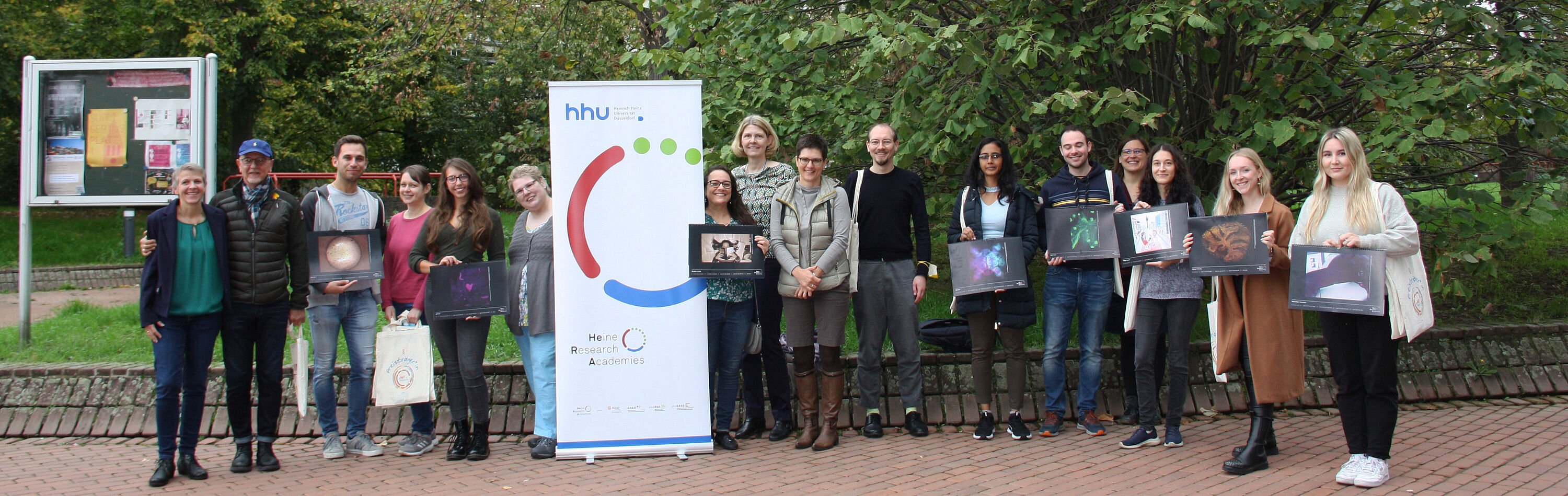
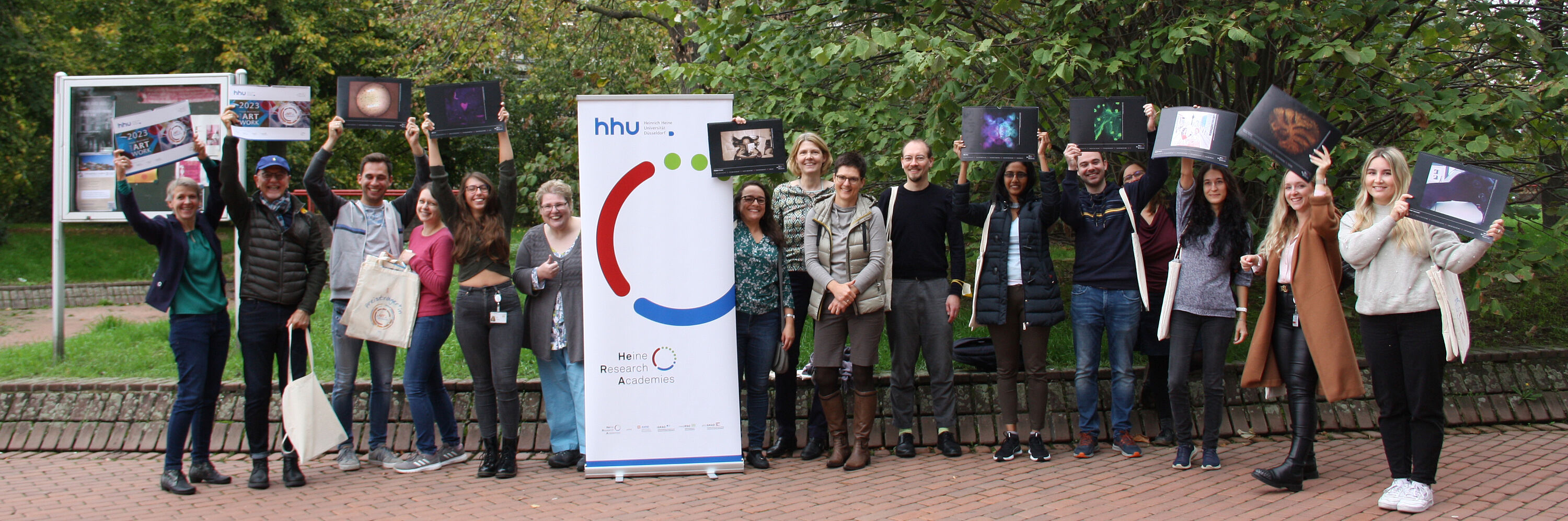
On 17.10.2022, the pART of Research Calendar 2023 was officially presented and the researchers commented on their images. Special guest at the event and inspiration for all artistically interested academics was HHU chemist Dr. Bernd M. Schmidt with his current collaboration project .
The calendar is now available at all officies of Heine Research Academies and the Diversity Section at the Heine Center for Sustainable Development - Diversity, Environment, Health.
The selected images of the 2023 pART of Research Calendar:
The Fun Journal of Nature and Science
Anay Kumar Maurya
Institute for Microbial Cell Biology, Faculty of Mathematics and Natural Sciences
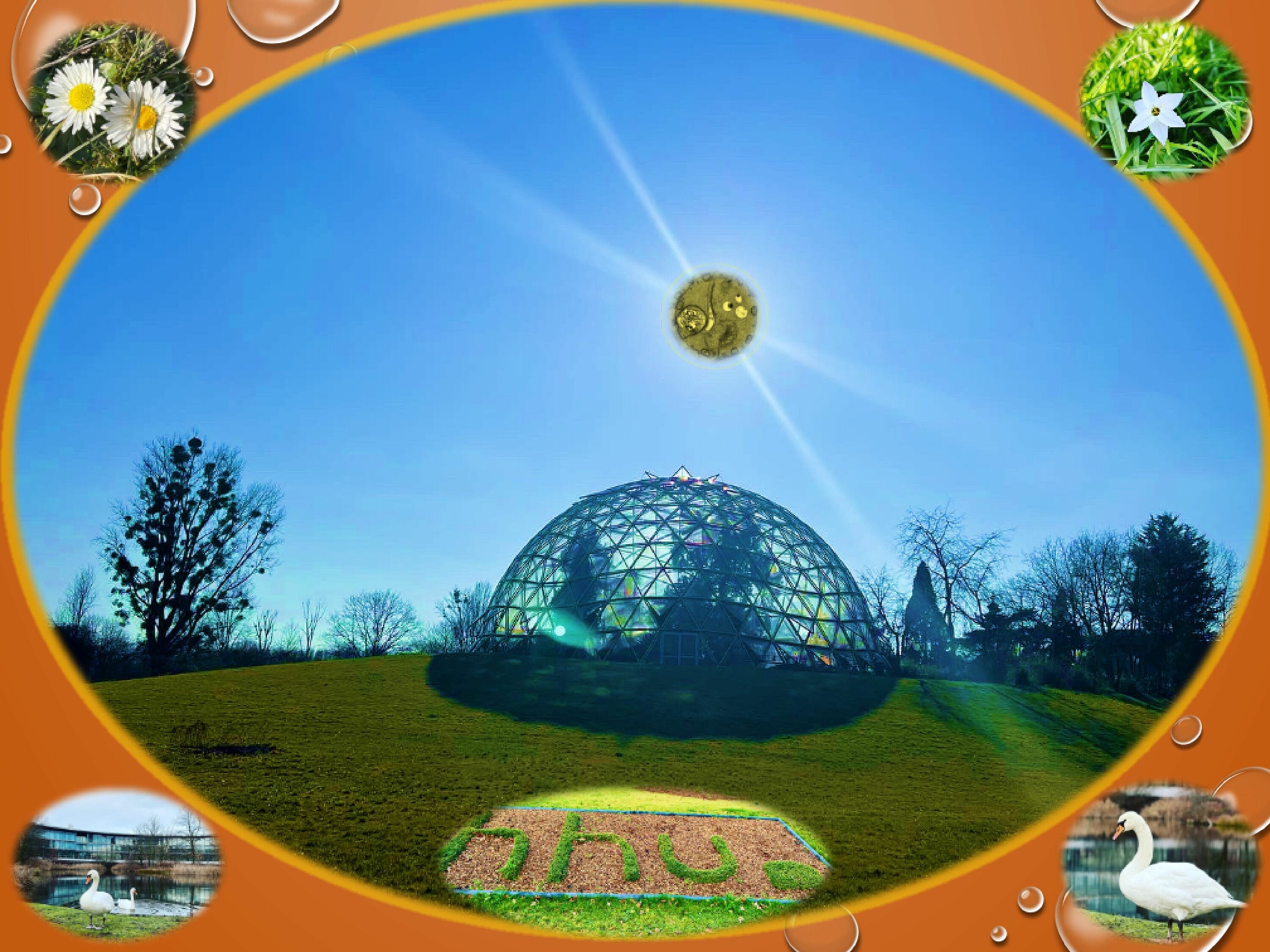
Transmission Electron Microscopy picture of Angomonas deanei ETP9 SKO (from Science) which replaces the Sun in the Botanical Garden Picture assembled with other pictures captured within the university (from Nature).
Spidermen at work
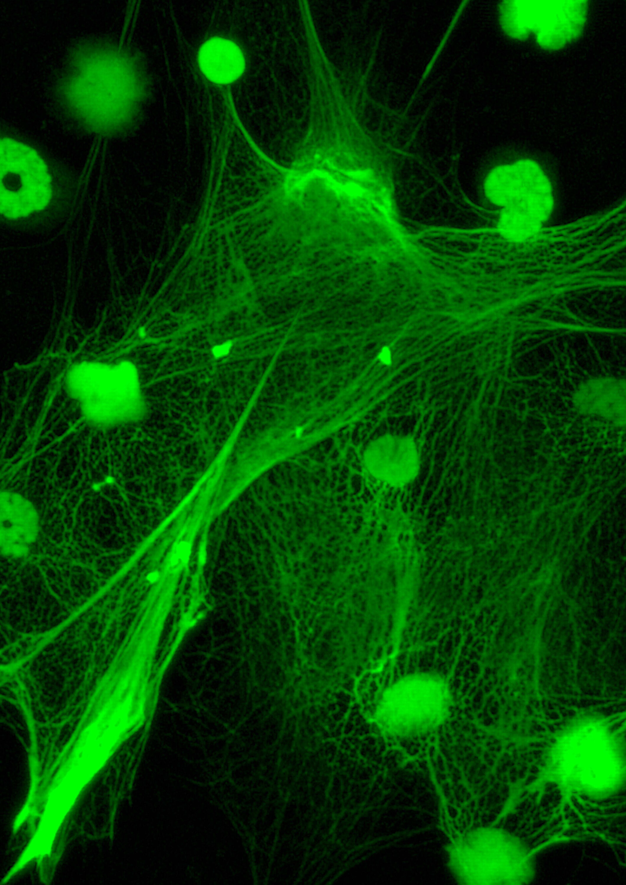
Marcel Marson
Institute of Medical Microbiology and Hospital Hygiene, (Doctoral Researcher at) Faculty of Mathematics and Natural Sciences
The formation of here shown neutrophil extracellular traps, short NETs, protect against infection, in particular against large pathogens. The nuclear chromatin of neutrophils decondenses into the cytoplasm, mix with cytoplasmic and granule components and expand into the extracellular space.
Asymmetry in Symmetry
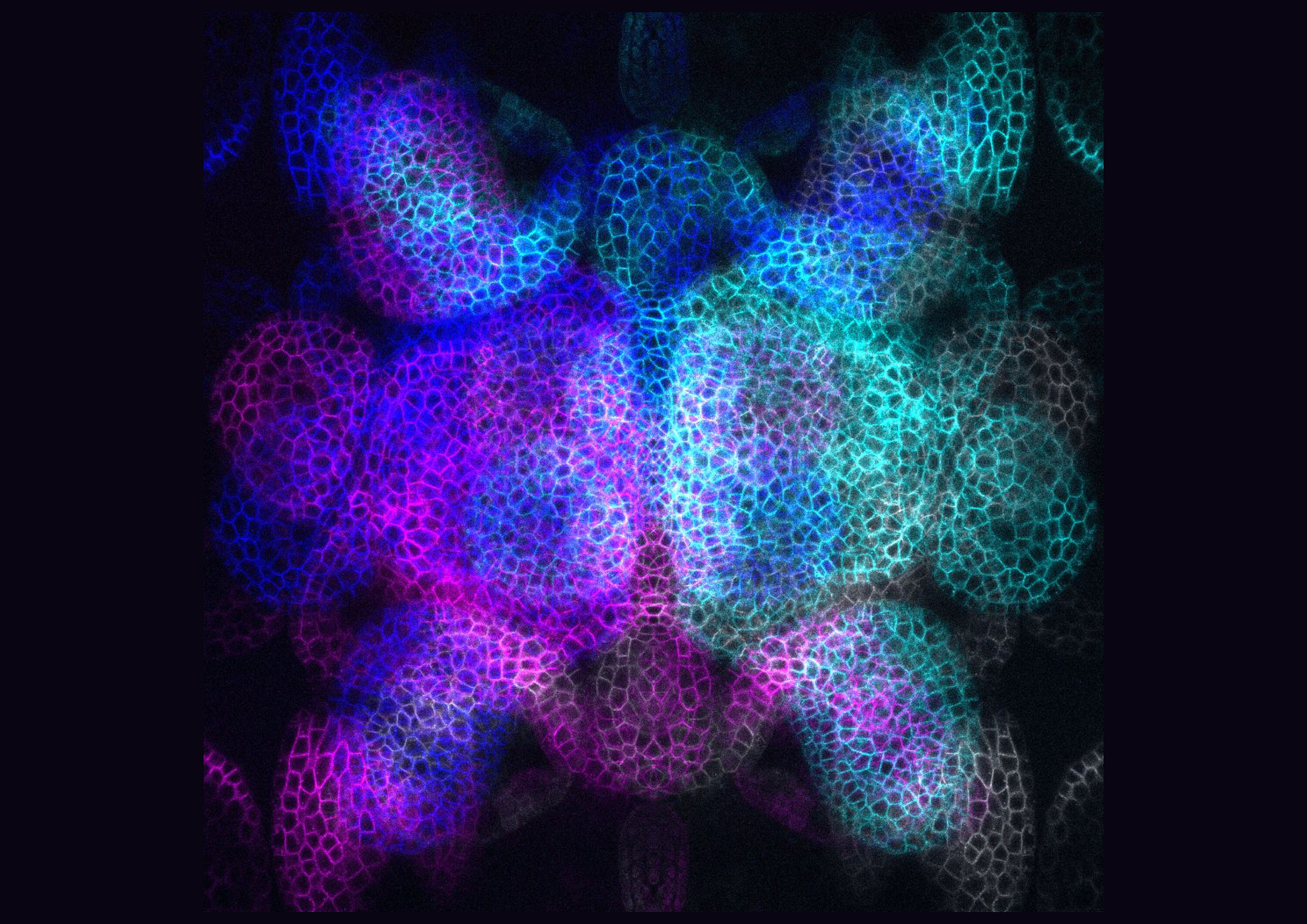
Dr. Madhumitha Narasimhan, Meik Henrik Thiele
Institute for Developmental Genetics , Faculty of Mathematics and Natural Sciences
This is an image of a plant meristem that I accidentally crushed. I transformed it into a symmetrical 3D pattern. I further added an asymmetrical colour palette upon it. Is it now symmetrical or asymmetrical? Perhaps symmetry is an illusion. Or is it a transient state from which asymmetry is born?
Myelin fibers of a mouse cerebellum
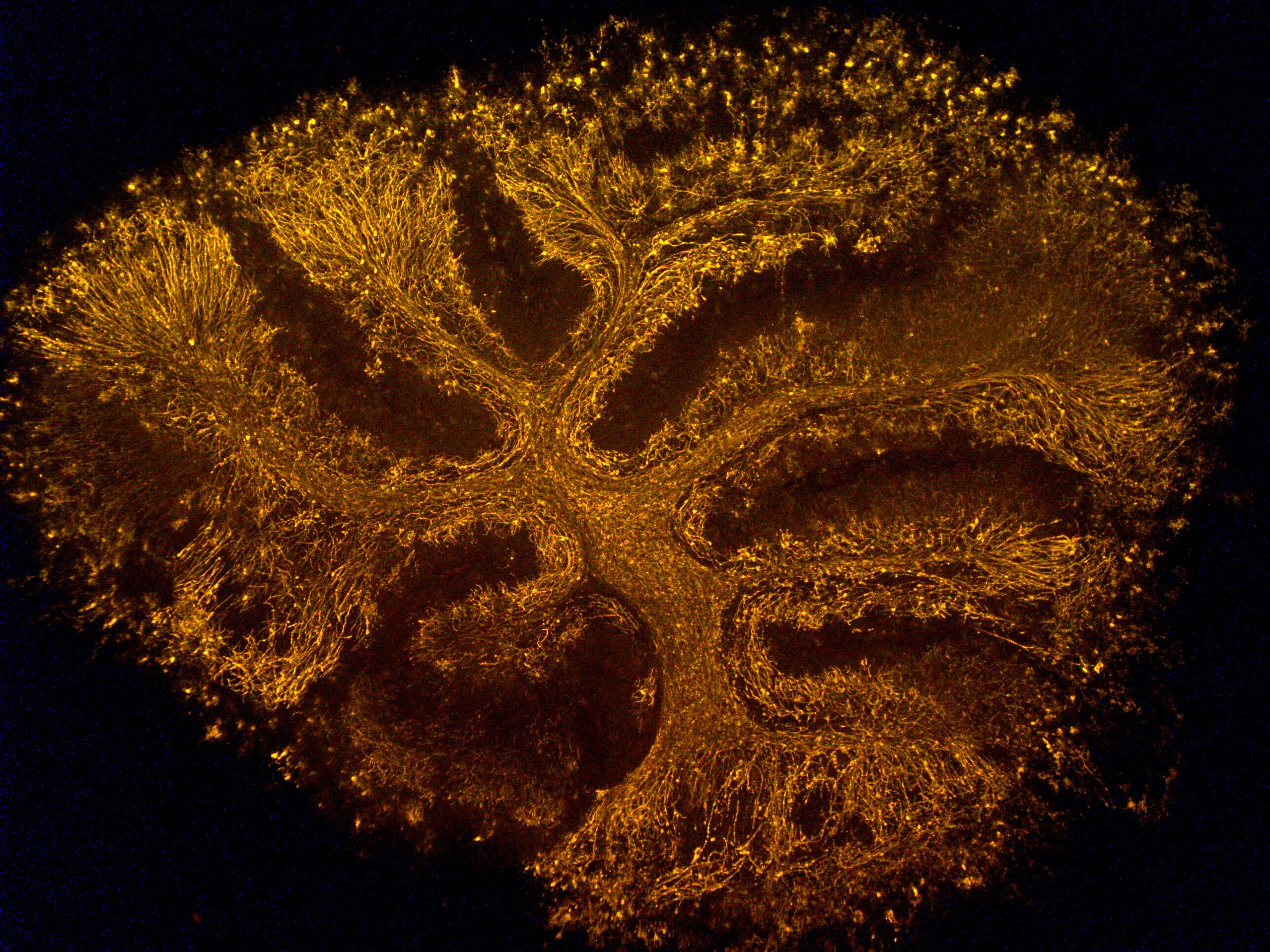
Leonie Thewes
Clinic for Neurology, (Doctoral Reseacher at) Faculty of Mathematics and Natural Sciences
The image shows the cerebellum of a mouse brain. In the experiment, the myelin fibers surrounding the neurons in the central nervous system were stained with an antibody. This allows visualization with the fluorescence microscope.
A Heart for Science

Verena Stehl, Maximilian Seidl, Anja Harbecke
Institute for Pathology, (Doctoral Researchers at) Faculty of Math. and Nat. Sciences
FFPE tissue in a confocal fluorescence microscope. Superficially deparaffinized tonsil tissue. Autofluorescence of the tissue was measured and a tonsil crypt formed a heart.
Love (for) Science
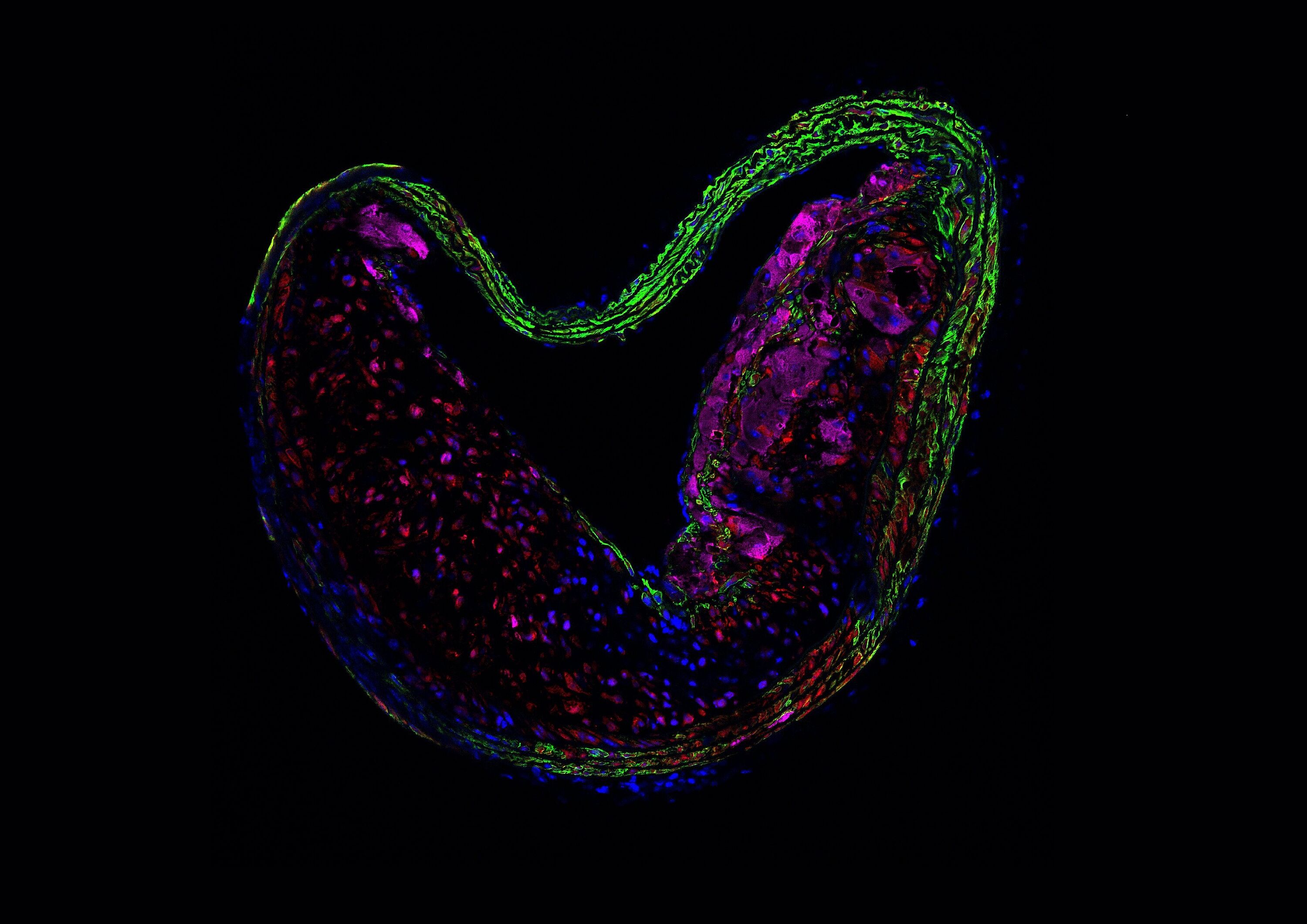
Fiona Cox
Institute for Translational Pharmacology, Medical Faculty
Shown is an immunofluorescence staining of a cross-section through a mouse blood vessel, specifically, the brachiocephalic artery (BCA), which has developed an atherosclerotic lesion or plaque as a result of an 18-weeks high-fat diet such that the lumen forms a heart.
Amyloid beta oligomer peaks
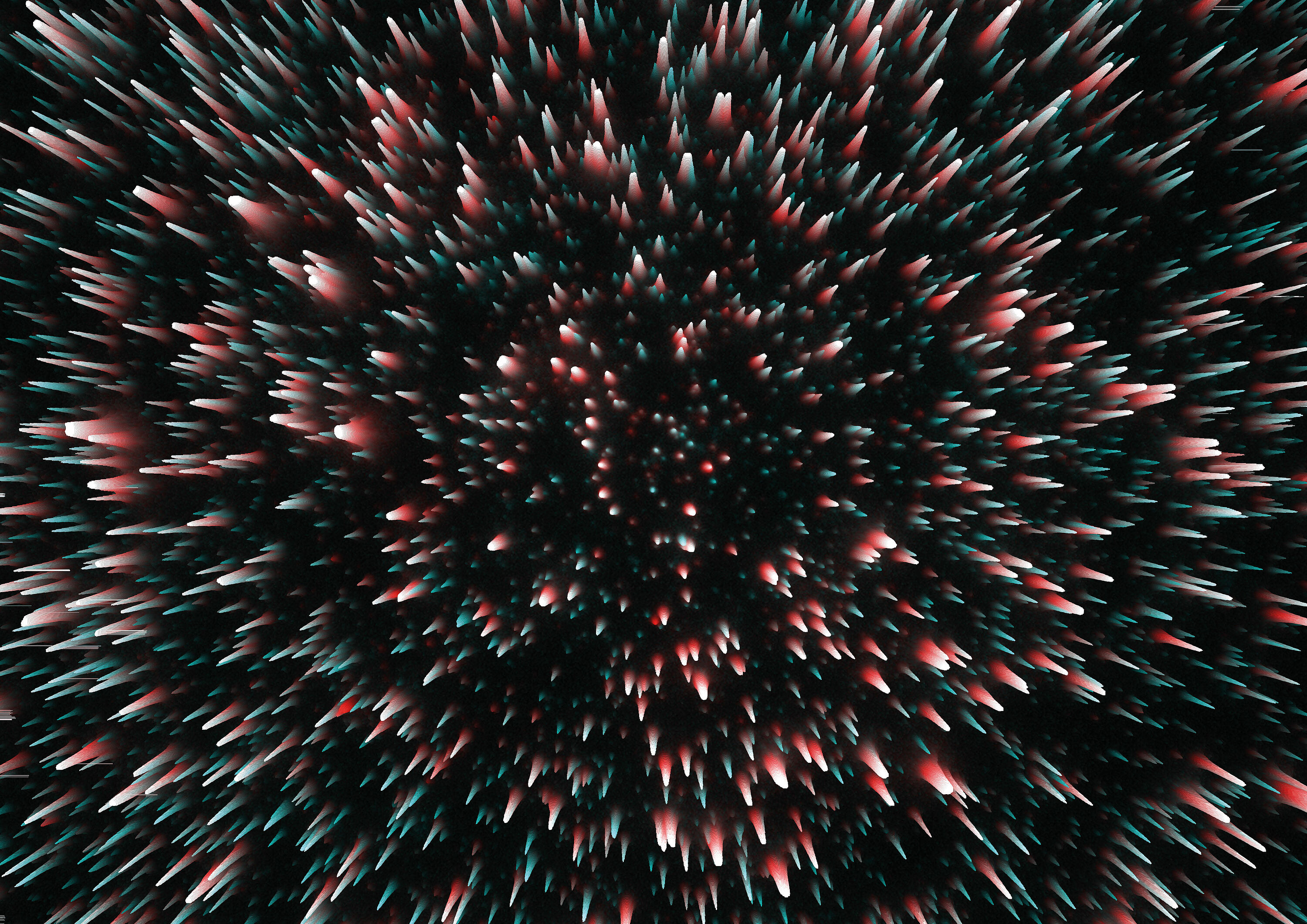
Bettina Kass
Institute for Physical Biology, Faculty of Math. and Nat. Scie.
Amyloid beta oligomer particles in brain homogenate from an Alzheimer’s disease patient, visualized as white-tipped peaks due to simultaneous binding of two different fluorophore-labeled antibodies. Based on a microscopy image ("sFIDA assay") with a total width of 114 µm.
Red Dot

Dr. Nadine Gier, Vita Zimmermann-Janssen, Regina Harms
Chair of Business Administration, esp. Marketing, Faculty of Business Administration and Economics
It's a little red dot, but it can shed (near-infrared) light on the darkness of the perception and processing of consumers. The picture shows an fNIRS device that is used in consumer neuroscience studies to measure neural activity.
Crystalline University
Dr. Mahsa Armaghan
Materials and Structural Research, Faculty of Mathematics and Natural Sciences
Crystalline university: brilliant ideas in crystalline university
Variety and diversity of life

Dr. Inga Mohr, Jannik Hornbergs , Christopher Endres
Institute of Botany, Faculty of Mathematics and Natural Sciences
Experiments lead to the desired result in the best case and sometimes simply to impressive works of art, typical pART of Research. Here you can see a variation of microorganisms grown on a mixture of nutrient media. Life is colorful, diverse and unstoppable!
Urban linguistic fieldwork in Düsseldorf
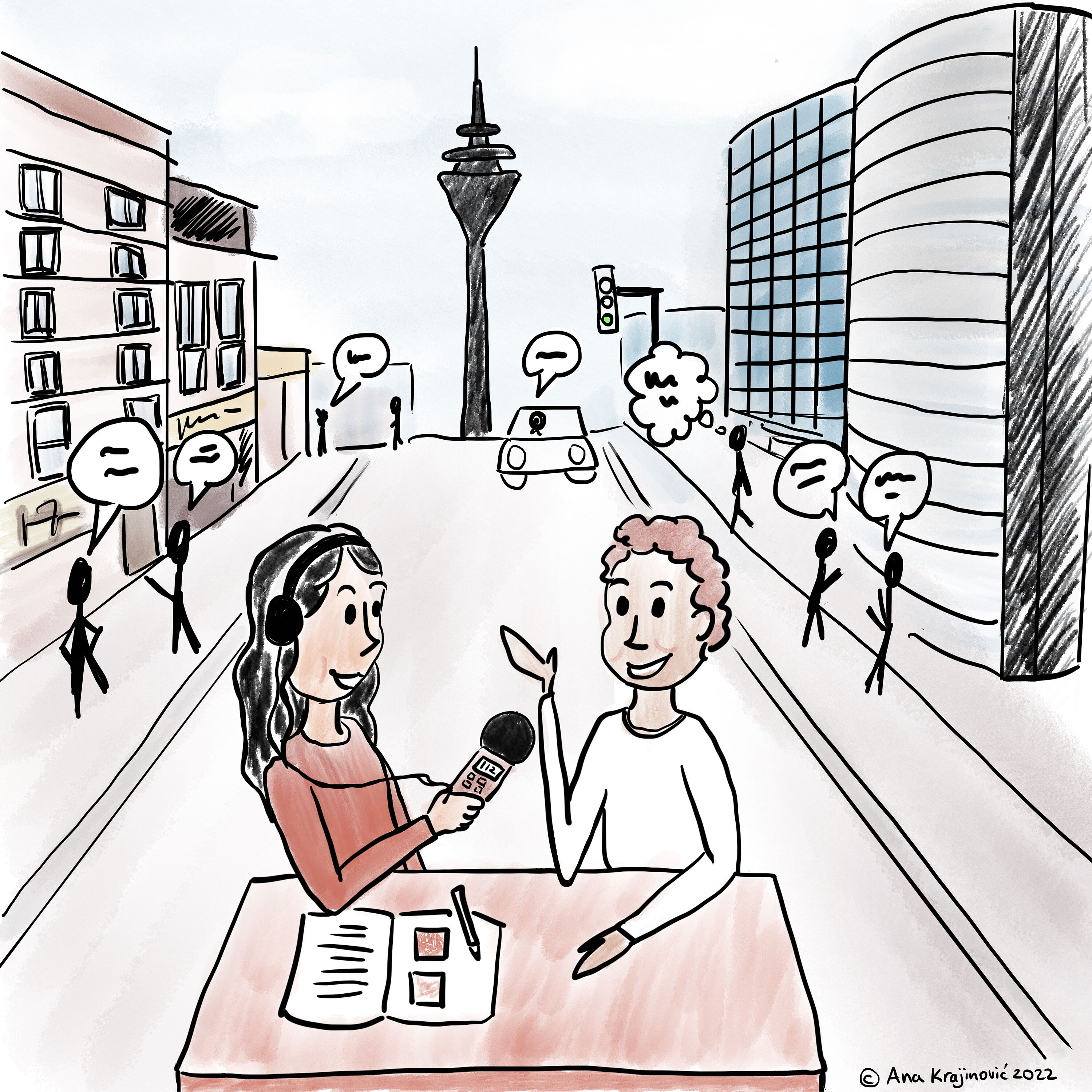
Dr. Ana Krajinović
Institute for Language and Information, Faculty of Arts and Humanities
With my colleagues (incl. Niklas Wiskandt and Isabella Greisinger), we teach linguistic fieldwork in a university course and put students in contact with speakers of lesser known languages spoken in Düsseldorf, such as Pontic Greek, Kannada, and Telugu.
Instagram
Pirahã Language

Juliana Neves-Müller
Institute of Language and Information, Faculty of Arts and Humanities
The language of the Pirahã Indians in the Amazon in Brazil poses a mystery to researchers, since it is very different from all other languages, including that of other Amazon Indians.