
Congratulations to the winners!
pART of Research Calendar Competition 2024
There is a lot of work in science - and also a lot of art. Whether it’s the coffee cup print on important notes, a picture of the last field trip or impurities under the microscope - surprises, mishaps, or the long-awaited result – every research project of yours makes a difference!
Under the motto: "Your Research - Your Art Work", 31 (!) pictures from four of the five faculties were submitted to the competition this year. We would like to thank all participants for their submissions .
Over 2800 (!!) people voted and selected for the 12 images which will be printed in the pART of Research calendar 2024. The calender will be available from Heine Research Academies this fall.
Official presentation of the pARTofResearch calendar 2024

© HeRA/HHU
On 24 October 2023, the pART of Research Calendar 2024 was officially presented. The award winners have reported impressive background information on their pictures and are happy about the 2024 calendar.
Here are the 12 winning images,
which will be printed in the calendar 2024!
jin jiyan azadi - Woman Life Freedom
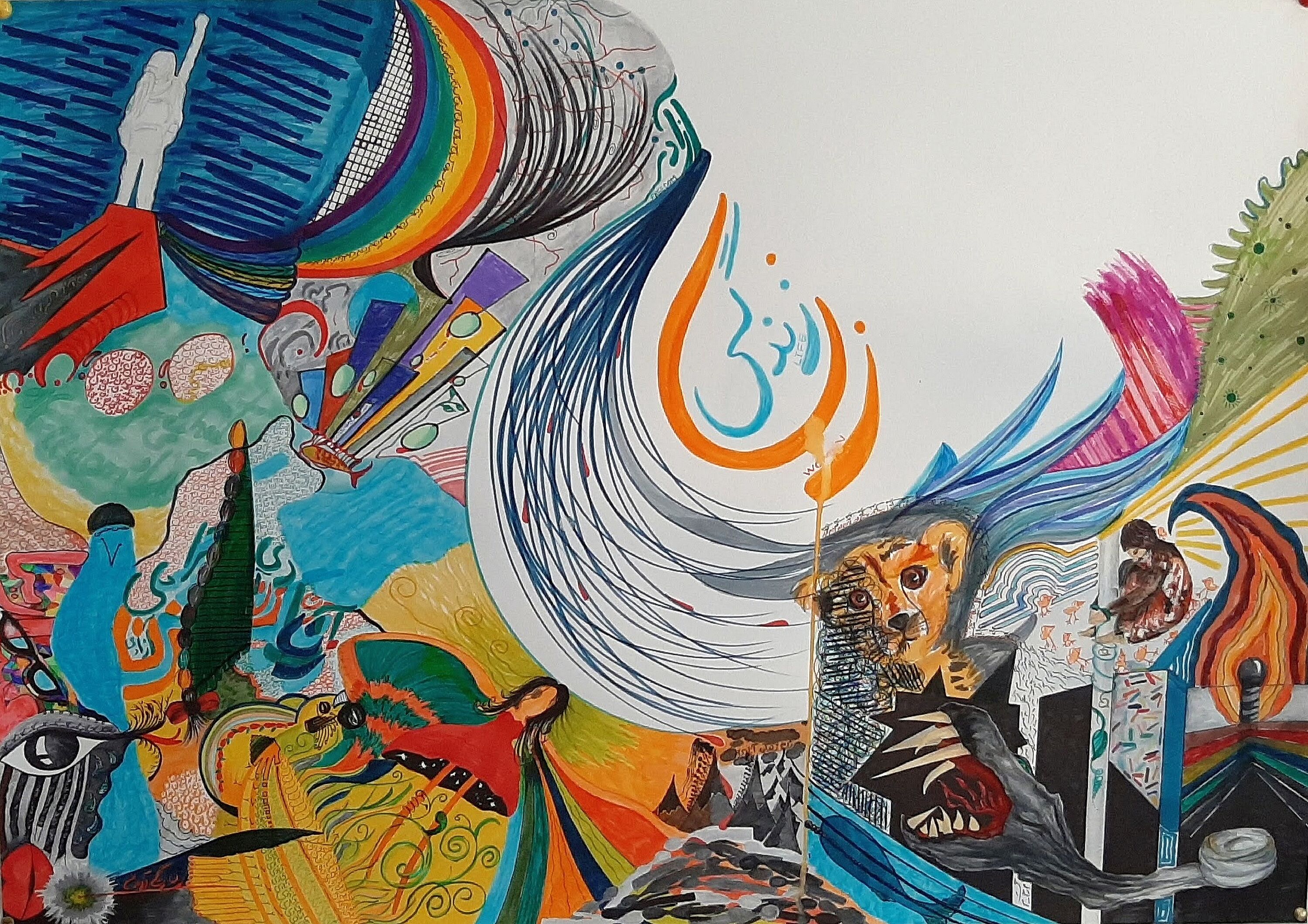
Sara Ipakchi
Philosophy VI, Faculty of Arts and Humanities
Sometimes unexpected things happen. They affect your work, your routine, your whole life. Then you think: Who am I?
The painting shows my ups and downs in relation to what has been happening in Iran since September 2022. The end of this madness will be the end of my painting.
Language portrait and living multilingualism
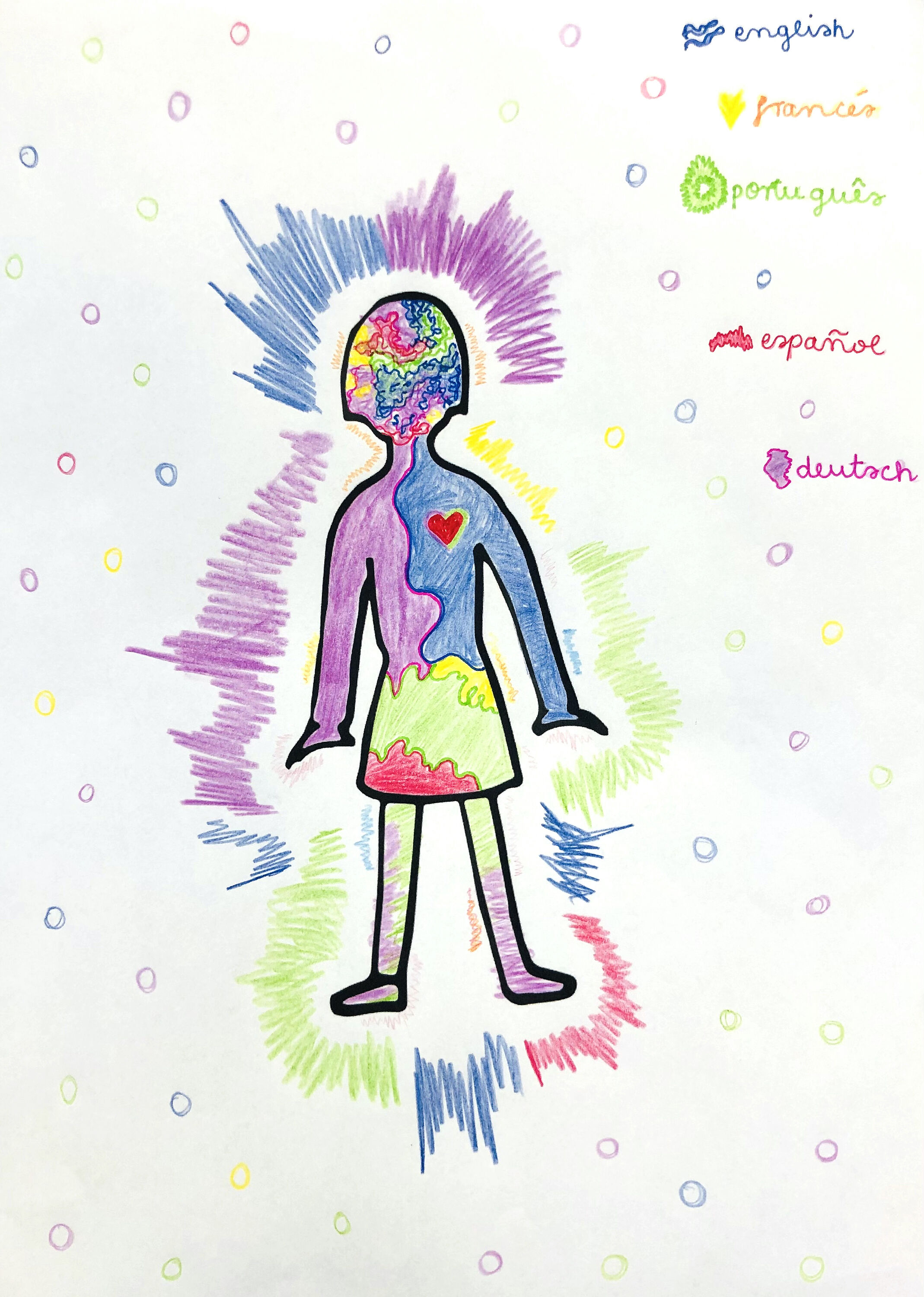
Juliana Neves-Müller
Institute of Language and Information, Faculty of Arts and Humanities
The language portrait method helps to analyze language practices of immigrants in Germany. It can generate valuable data on how multilingual people construct their own identity and repertoire in a new country.
Rainbow in a dish
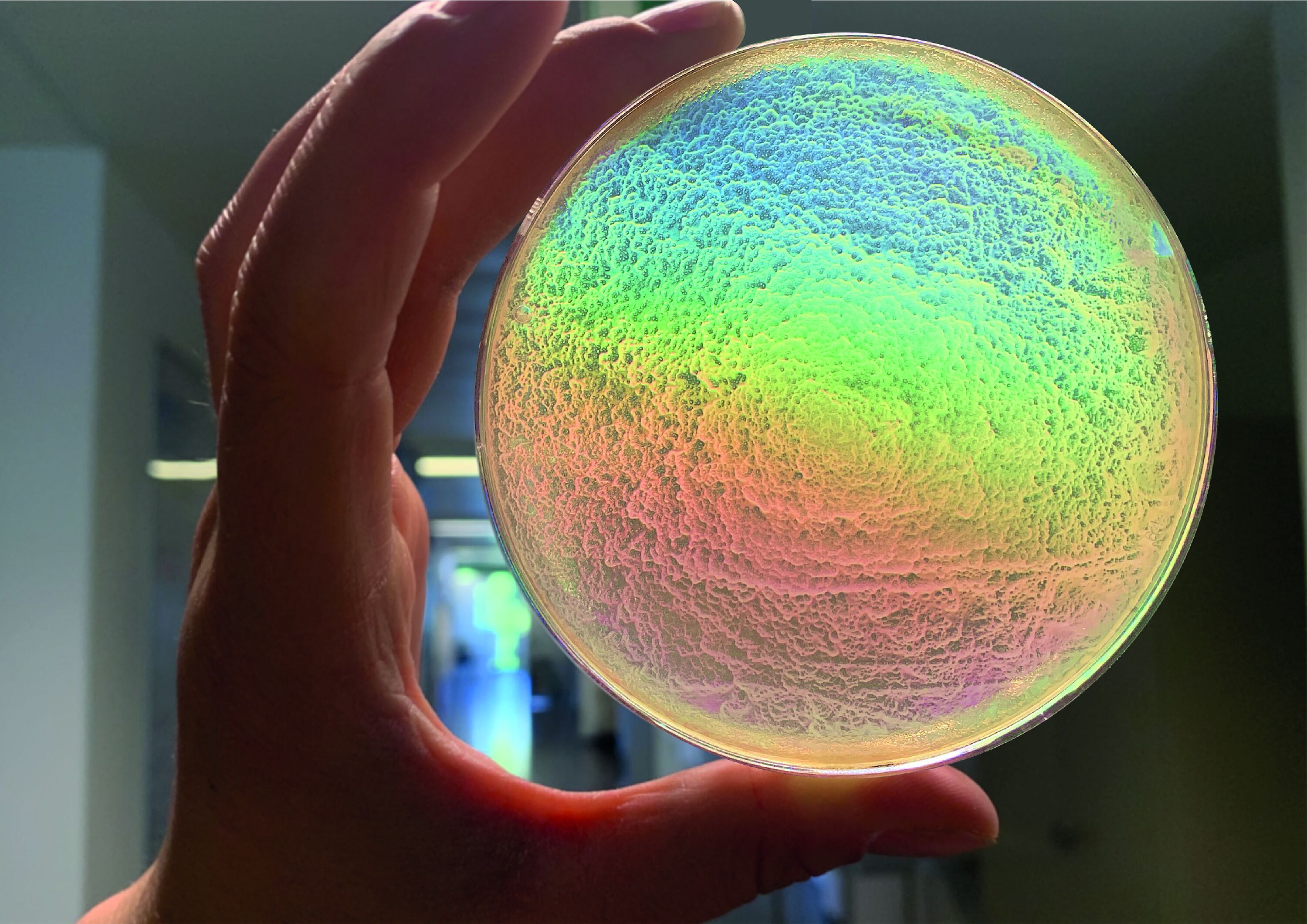
Luzie Kruse, Dr. Stephan Thies
Institute for Molecular Enzym Technology, Faculty of Mathematics and Natural Sciences
Imagine, it's early in the morning, you're tired and not expecting anything special, but then you open the cultivator, get your plates and your bacteria amaze you by beautifully iridescing in the sunlight. Sometimes science can be surprisingly artful and gratifying in miraculous ways.
Host control over its bacterium

Anay Kumar Maurya, Georg Ehret, Miriam Bäumers and Prof. Dr. Eva Nowack
Institute for Microbial Cell Biology, Faculty of Mathematics and Natural Sciences
Angomonas deanei (a trypanosomatid), harbors a single bacterium in its cytoplasm. Endosymbiont-targeted protein 9 (ETP9), a host-encoded protein, localizes at the bacterial division site. Heterozygous knock outs of ETP9 show aberrant division phenotypes, e.g., the formation of bacterial chains.
Cosmic expansion in a nutshell
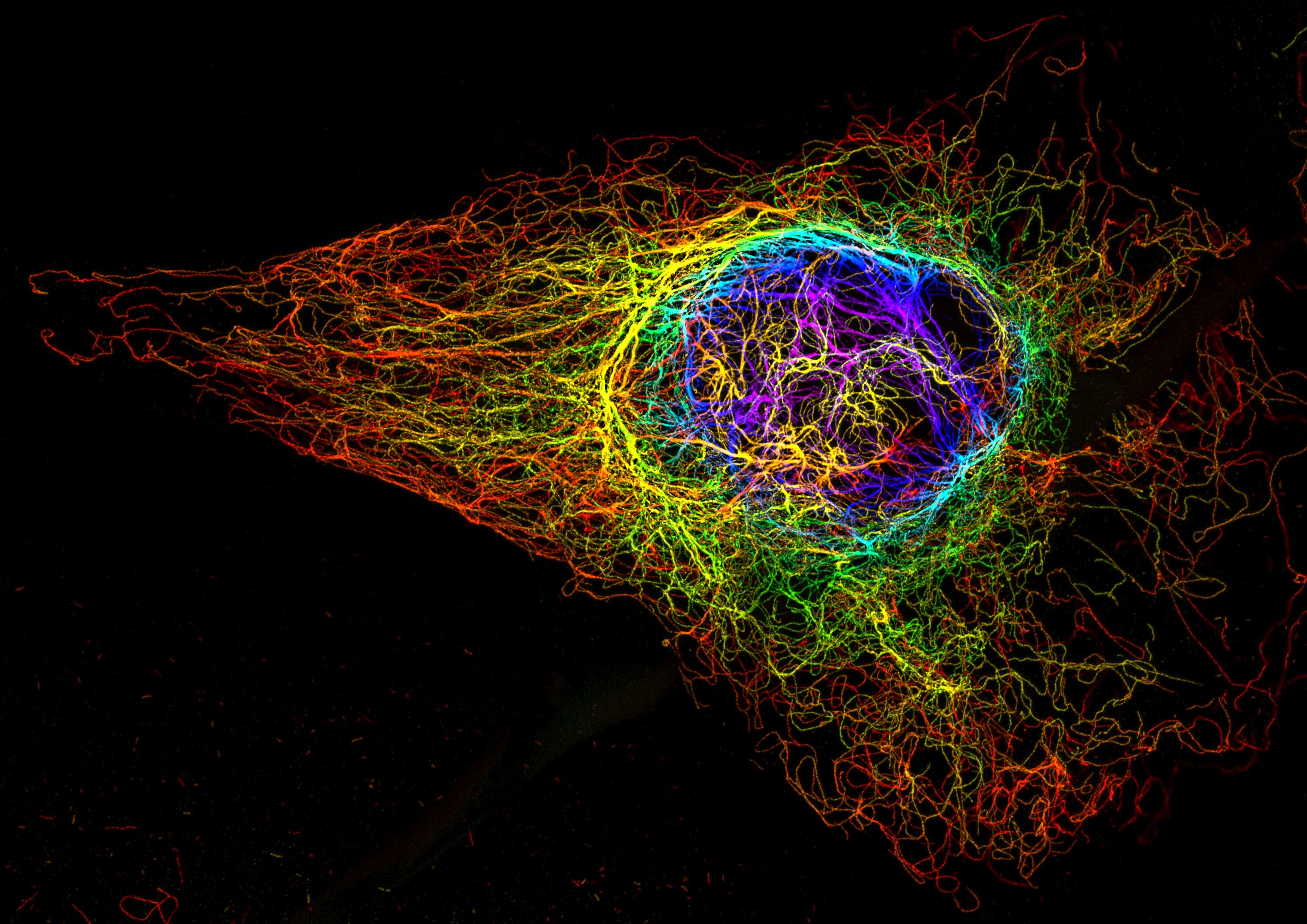
Dr. Sebastian Hänsch
CAi - Center for Advanced Imaging, Faculty of Mathematics and Natural Sciences
Vimentin cytoskeleton of a human cell, which was expanded ~4x by sample preparation. Combined with confocal fluorescence microscopy, expansion microscopy reveals finest details of the filaments, while the overall structure resembles the cosmic matter network. The largest among the smallest!
Moonlight
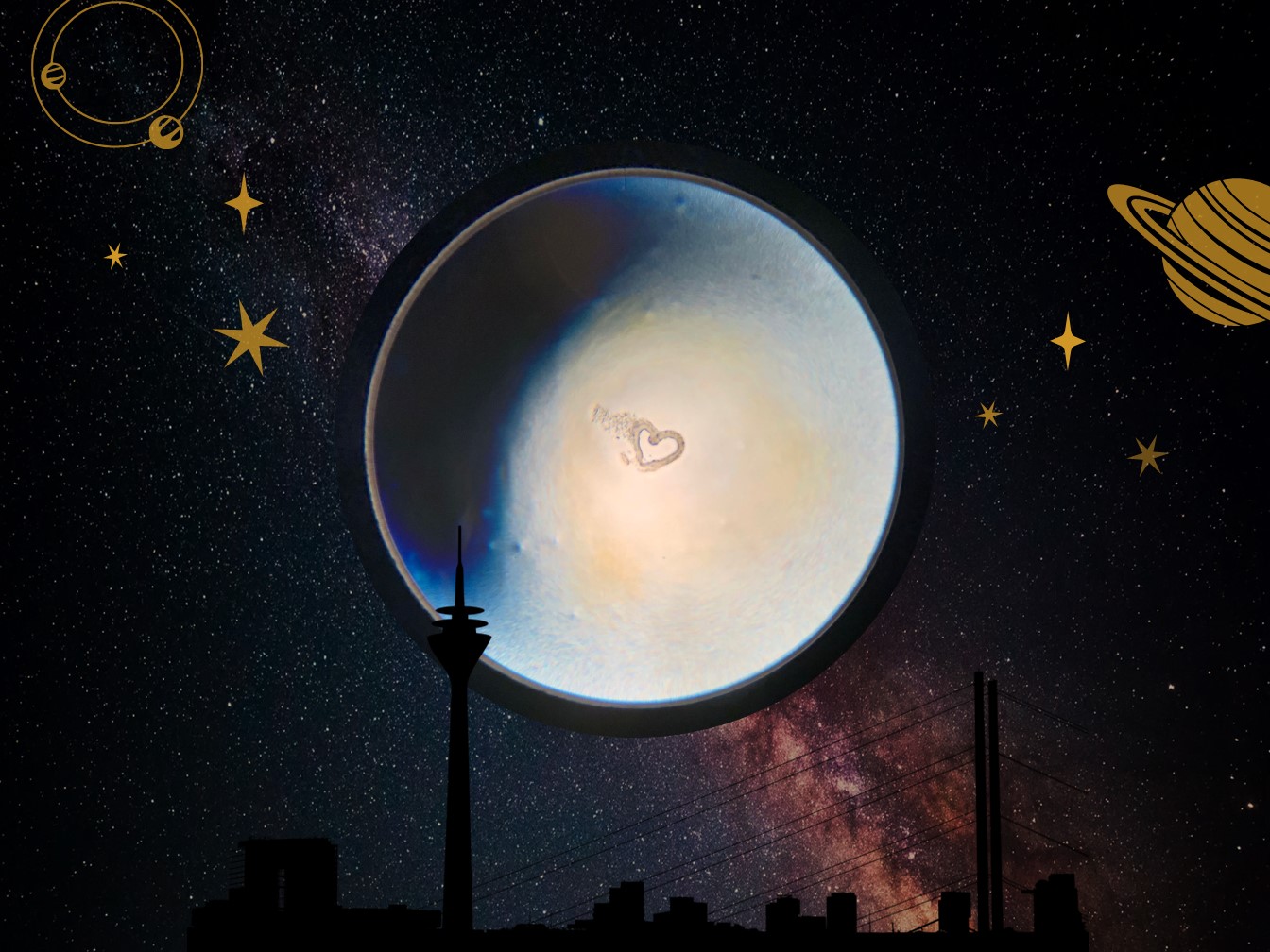
Anika Prinz
Institute for Translational Pharmacology, Medical Faculty
I took a snapshot of a cross section through a murine blood vessel. It was crushed during sectioning, thus heart-shaped, and I couldn‘t use it for further analysis.
It being mistaken for a picture of the moon later on gave rise to a new purpose and I transformed it into this (p)ARTwork of Research.
I(D)ris
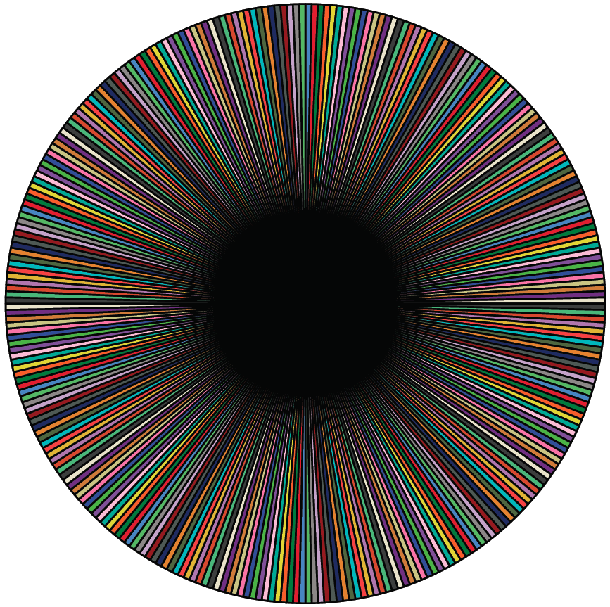
Dr. Nadine Gier, Vita Zimmermann-Janssen; Regina Harms
Chair of Business Administration, esp. Marketing, Faculty of Business Administration and Economics
Why should you plot the participants' IDs as a piechart? - Because it looks fantastic! It's like looking into the "eye of your research sample".
Infinity Mirror Molecule
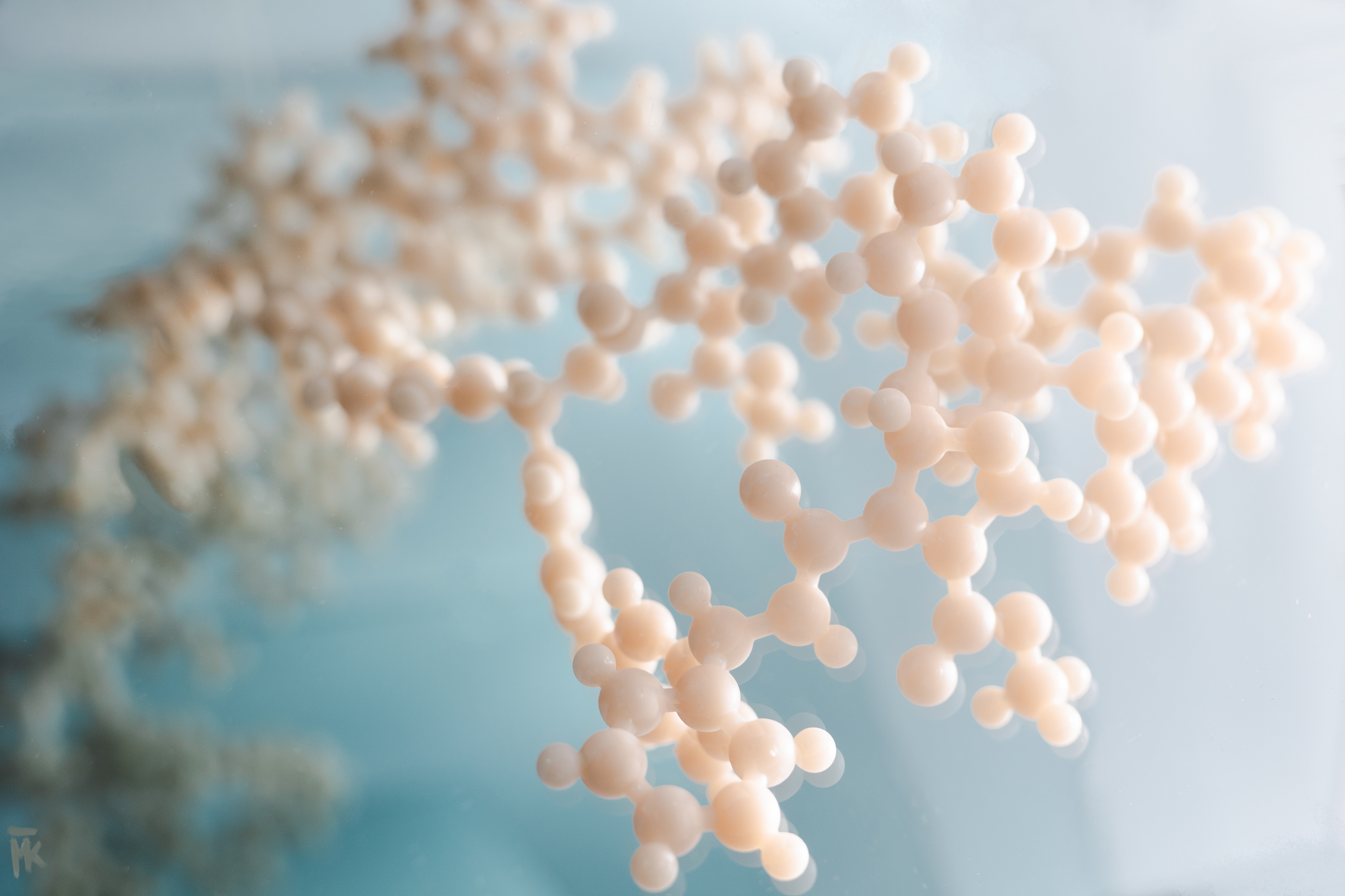
Moritz Klischan
Institute of Bioorganic Chemistry, Faculty of Mathematics and Natural Sciences
This image shows a 3D-printed molecule obtained as an X-ray crystal structure during a research stay at the University of Toronto. The molecule was replicated with mirrors to depict the crystal macroscopically.
University Trees
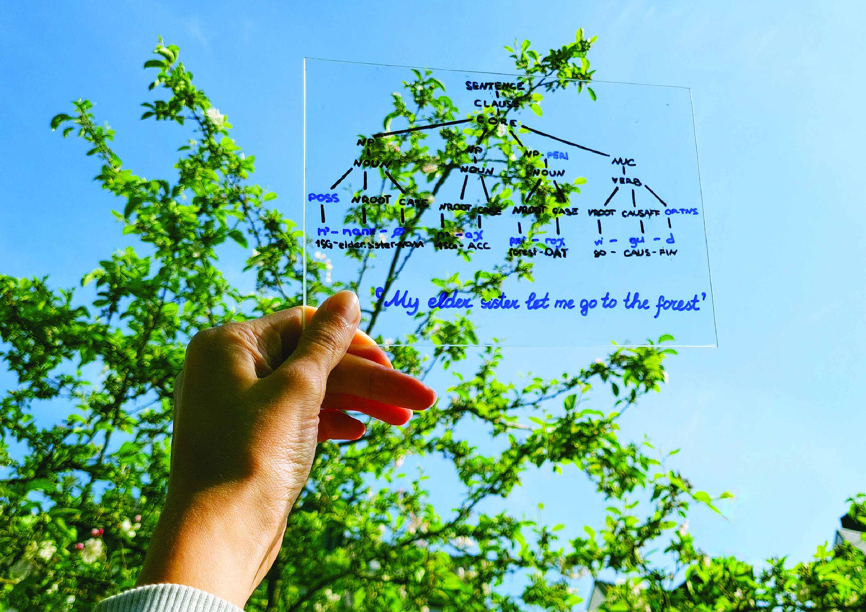
Valeria Generalova
Institute for Linguistics and Information Science, Faculty of Arts and Humanities
Dr. Daria Kohler, KU Leuven
In linguistics, we have so many trees that sometimes can't see the forest for them.
If so, it's time to take a breath of fresh air.
This sentence in Nivkh — a language spoken on Sakhalin Island by ca. 200 people — is an invitation: to a forest, a research, a fairytale.
SEM image of the tail of C. elegans
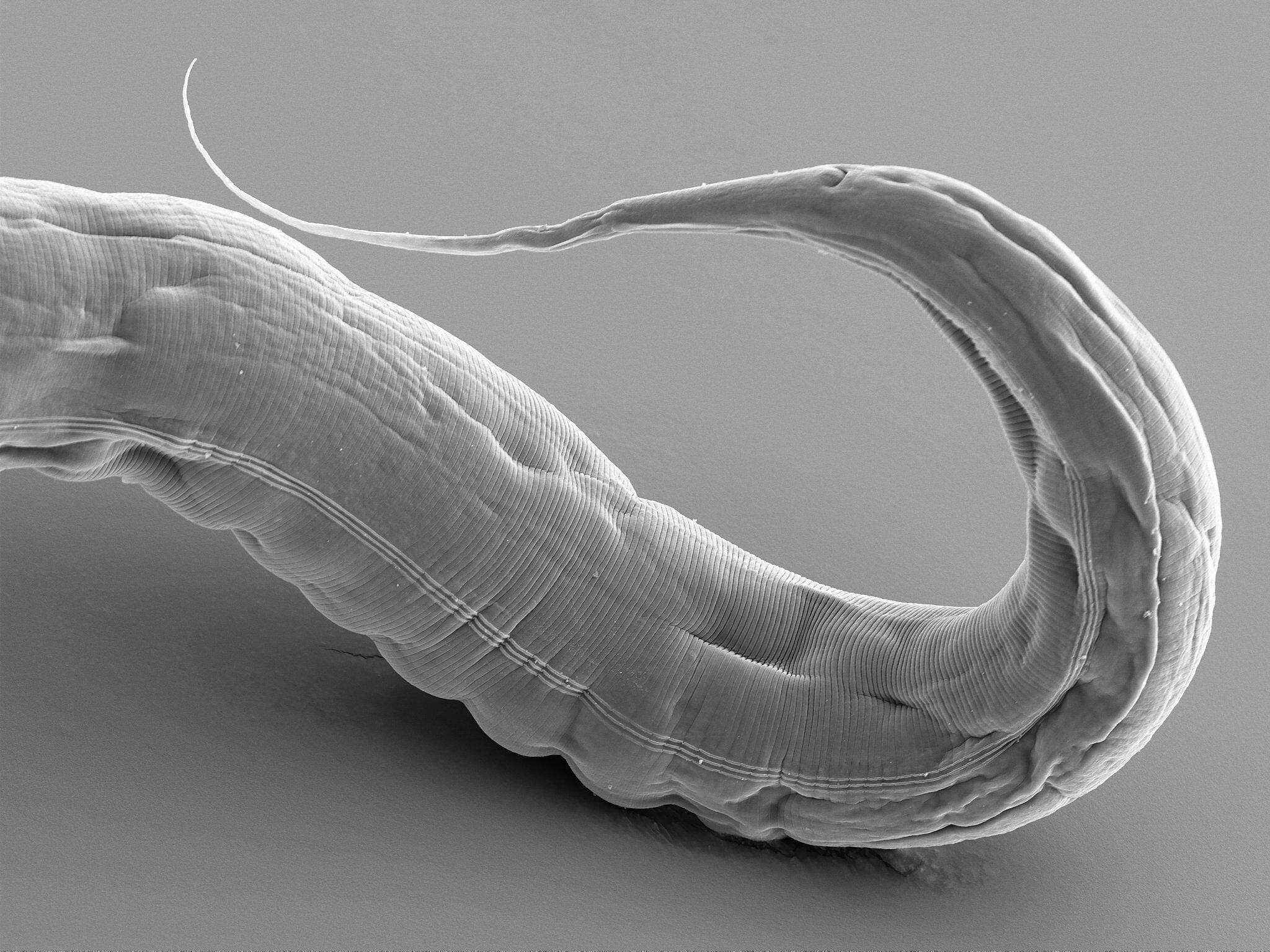
Alexandra Daum, Steffen Köhler
Institute of Cell Biology, Faculty of Mathematics and Natural Sciences
The image shows the tail of the nematode Caenorhabditis elegans and was created with a scanning electron microscope. With this unique method, we were able to precisely visualize the cuticle of the hermaphrodite.
Upside Down Moods
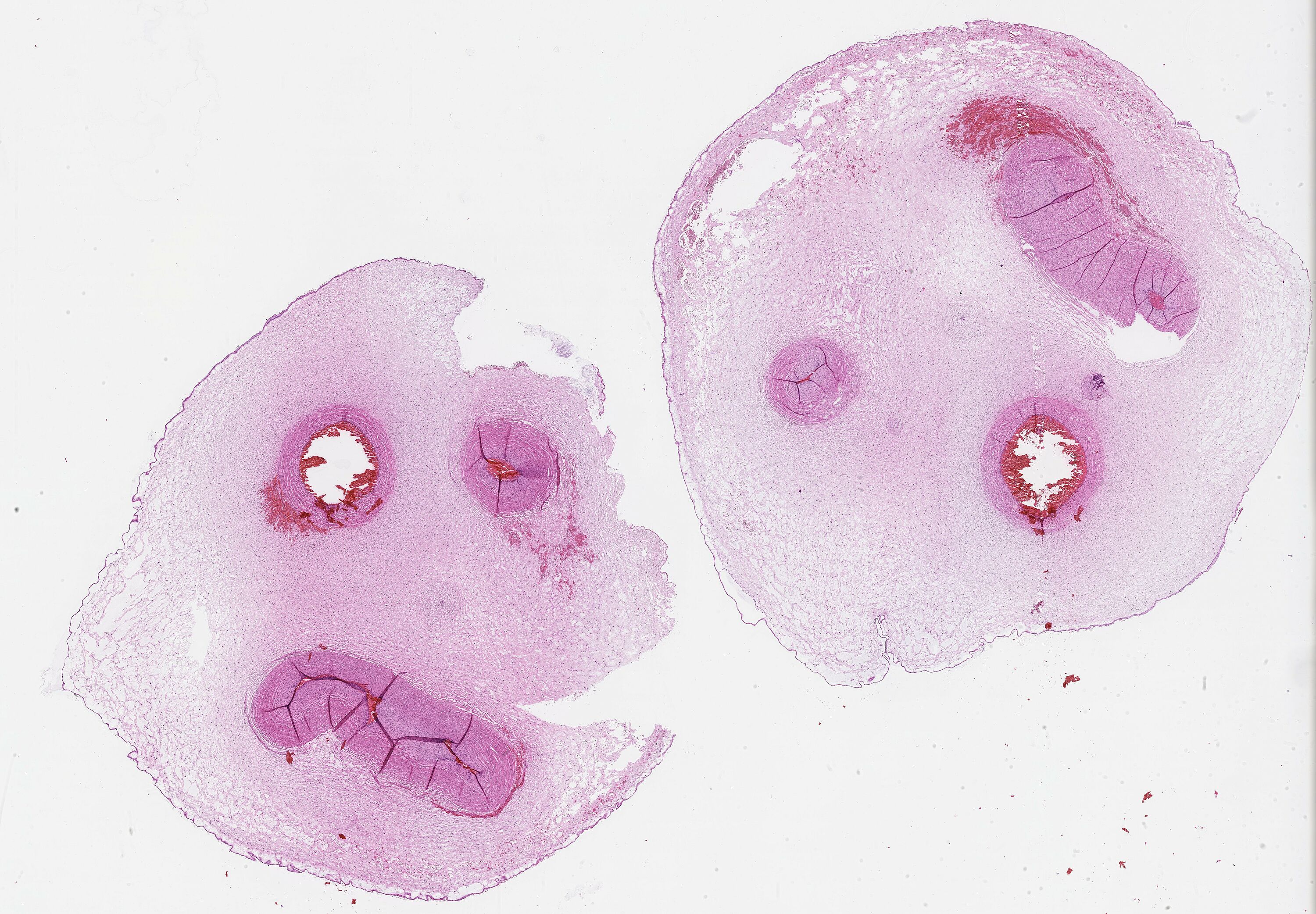
Verena Stehl, Muhannad Al Kallaa
Institute for Pathology, Doctoral researcher of the Faculy of Mathematics and Natural Sciences
In histology, there are good specimens as well as bad ones. Our image represents the whole thing with the smileys formed by the specimen of an umbilical cord in the HE section.
A red dot on the trail

Lukas Theissen, Dr. Christopher Nelke
Department of Neurology, Medical Faculty
A muscle section is seen by immunofluorescence staining. Laminin-Beta-1 marks the cell membrane of each muscle cell in green. A single nucleus stains positive for p21 in red - a marker for cellular senescence.